Imaging, anatomy and shape
Turning clinical images into anatomical models: segmentation, meshing, fibre architecture and statistical shape analysis, at the scale of whole cohorts rather than single cases.
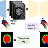
Turning clinical images into anatomical models: segmentation, meshing, fibre architecture and statistical shape analysis, at the scale of whole cohorts rather than single cases.
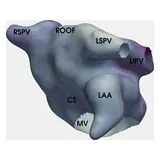
Mechanisms that start and sustain atrial fibrillation, which patients an ablation will help, and how it can be targeted for a particular atrium rather than a generic one.
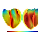
Why dangerous ventricular rhythms arise in scarred and remodelled hearts, which regions of a particular heart can sustain them, and where an ablation should be aimed.
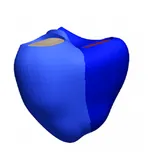
Models tied to one person's measurements, built for many people at once, so that questions can be asked of a population of hearts rather than a single example.
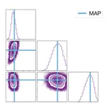
How far a model's predictions can be trusted: emulators, sensitivity analysis and calibration that state what the data can identify, and verification of the simulators themselves.
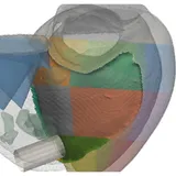
Electromechanical models of the failing and hypertrophied heart, and how pacing, resynchronisation and other treatments can be planned for a particular patient.
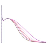
Biophysical models of the myocyte, from ion channels to force generation, applied to drug action and safety, to sex hormones, and to carrying results from the laboratory to patients.
Networks of cardiovascular digital twins, aimed first at pulmonary arterial hypertension, a condition with high mortality and frequent worsening between clinic visits.
A centre for model-informed medicine, applying mechanistic and data-driven modelling to the development of drugs, devices and lifestyle interventions.