Imaging, anatomy and shape
Turning clinical images into anatomical models: segmentation, meshing, fibre architecture and statistical shape analysis, at the scale of whole cohorts rather than single cases.
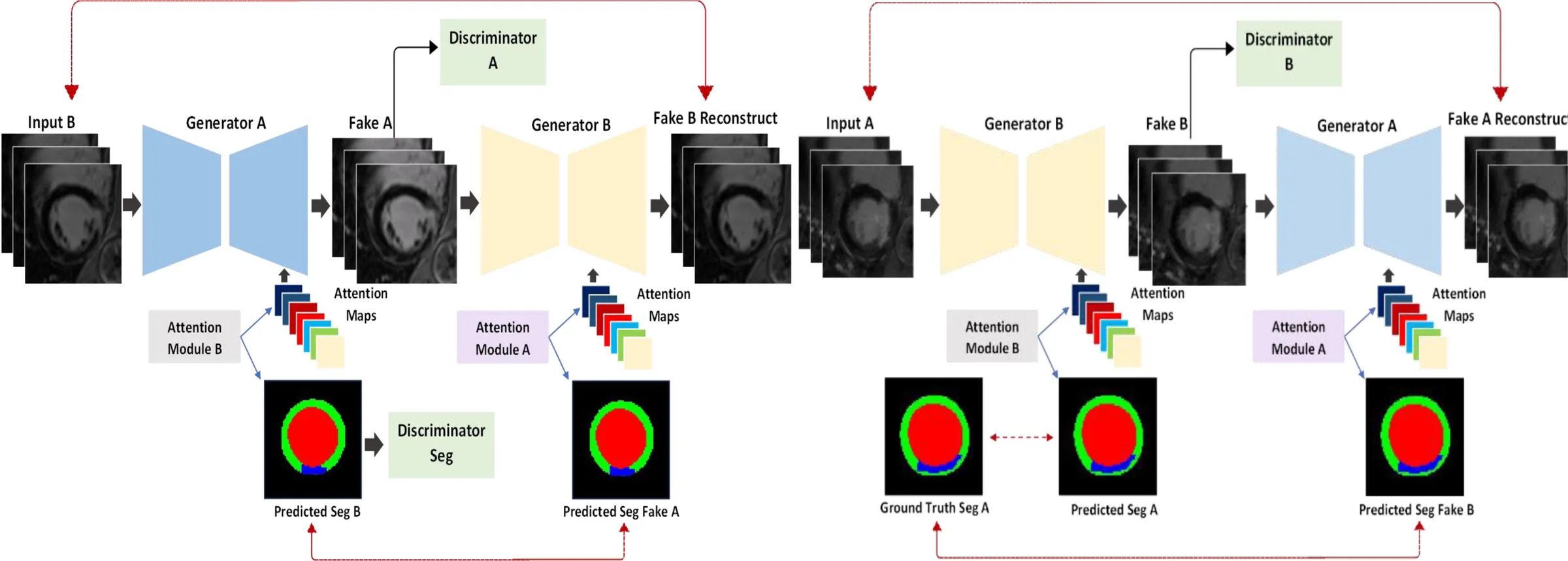
Clinical images are turned into anatomical models: the chambers segmented, the surfaces reconstructed, the myocardium meshed, and fibre orientation assigned. Deep learning does much of this, from segmentation networks for cardiac MRI and CT to networks that reconstruct the shape of the heart from cine MRI or from a few standard views.
Statistical shape analysis then describes how anatomy varies across a population, and how that variation changes the function a model simulates. A shape model can also describe the uncertainty in one patient’s anatomy, and how that uncertainty changes the arrhythmias the model predicts.
Recent papers
AortaST: A Student-Teacher Framework for Multi-class Aortic Segmentation
Integrating Imaging and Invasive Pressure Data into a Multiscale Whole-Heart Model
Influence of Oestrogen on Atrial Fibrillation Risk: An In-Silico Study
Is regional atrial strain a useful surrogate of regional atrial fibrosis in atrial cardiomyopathy?