Models and data
Four-chamber meshes from hypertrophic cardiomyopathy patients
2026
Four-chamber tetrahedral meshes of five patients with hypertrophic cardiomyopathy, one for each anatomical phenotype (mid-to-apical, isolated basal and milder asymmetric left ventricular hypertrophy, left ventricular outflow tract obstruction, and an undifferentiated pattern), with element tags and fibre directions, built with the CEMRG HeartBuilder pipeline. Four further records hold the simulations run on them: reaction-eikonal activation-time maps, passive inflations of four of them, the unloaded reference configurations, and closed-loop cardiac cycles coupled to the CircAdapt circulation, with the inputs and outputs of the global sensitivity analyses.
Rodero C, Curran L, Lanyon CW, Inglese P, Qayyum A, et al. (2026) Computational models show that functional remodeling, not anatomy, reshapes physiological determinants of cardiac performance in hypertrophic cardiomyopathy. J Mol Cell Cardiol 218: 137-150
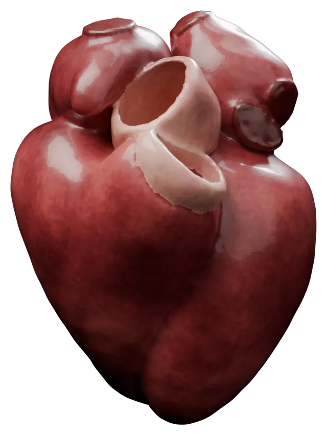
Drag to turn in 3D
Case HCM4 of the five four-chamber meshes, rendered from the published data, which are shared under CC BY 4.0. A model for the human fetal ventricular myocyte electrophysiology
2026
A model of human fetal ventricular myocyte electrophysiology at ten weeks of gestation. It includes the fetal-specific T-type calcium and funny currents, and is calibrated to mRNA expression data and experimental measurements.
Avezzù A, Longobardi S, Alvarez-Laviada A, Schultz F, Gorelik J, Williamson C, Niederer SA. A model for the human fetal ventricular myocyte electrophysiology. PLoS Comput Biol. 2026 Jan 27;22(1):e1013889.
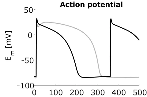
Action potential of the fetal model at a cycle length of 353 ms, a heart rate of 170 beats per minute at ten weeks of gestation (black), against the adult model of ten Tusscher and Panfilov on which it is built (grey). The first panel of Figure 9 of Avezzù et al. (2026), PLoS Computational Biology, under CC BY 4.0. Sex- and Disease-Stratified Patient-Specific Four-Chamber Heart Models from Clinical CT
2026
Fifty (50) patient-specific four-chamber heart models derived from clinical CT scans, balanced by sex (50% female) and near-balanced by condition. Each model provides anatomically detailed tetrahedral meshes with fibre orientations plus parameter files for benchmark electrophysiology and passive inflation simulations.
Solís-Lemus, J. A., Barrows, R. K., Rodero, C., Strocchi, M., Montarello, N., Lahoti, N., ... & Niederer, S. A. (2026). Assessing the importance of sex and disease-specific anatomy in electrophysiology and mechanical simulations with a newly developed public virtual cohort of four-chamber heart models. PLOS Computational Biology, 22(6), e1014325.
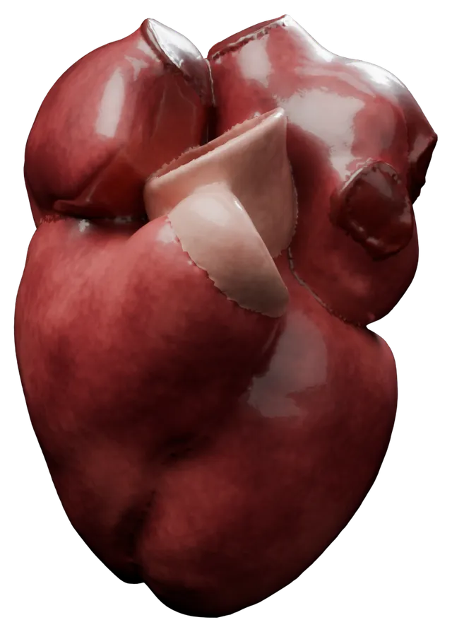
Drag to turn in 3D
Case CTW6MAJL of the 50 four-chamber models, rendered from the published data, which are shared under CC BY 4.0. Regional heterogeneity in left atrial stiffness impacts passive deformation in a cohort of patient-specific models
2025
Left atrial meshes built from end-diastolic CT of ten heart failure patients recruited for an upgrade to cardiac resynchronisation therapy, with patient-specific wall thickness, rule-based fibres and five regions from universal atrial coordinates, and meshes of the epicardial adipose tissue around the atria. The record also holds the training data of the Gaussian process emulators for each wave of history matching, the image-derived targets, and the Markov chain Monte Carlo samples of the calibration.
Baptiste TMG, Rodero C, Sillett CP, Strocchi M, Lanyon CW, et al. (2025) Regional heterogeneity in left atrial stiffness impacts passive deformation in a cohort of patient-specific models. PLoS Comput Biol 21(11): e1013656
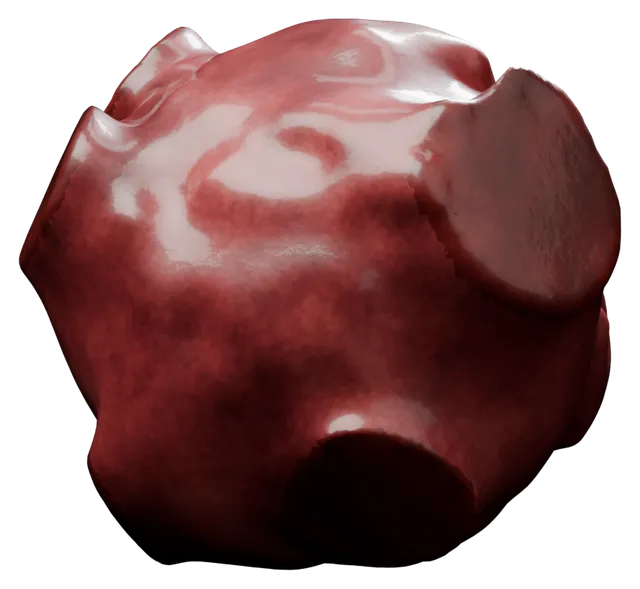
Drag to turn in 3D
Case 01 of the ten left atrial meshes, rendered from the published data, which are shared under CC BY 4.0. Cardiac digital twins at scale from MRI: Open tools and representative models from ~ 55000 UK Biobank participants - Segmentation Networks and Representative Meshes from UK Biobank
2025
We present a database of representative left and right ventricular meshes constructed from patient-specific models based on a large cohort of ~55k participants from UK Biobank.
Ugurlu D, Qian S, Fairweather E, Mauger C, Ruijsink B, Dal Toso L, Deng Y, Strocchi M, Razavi R, Young A, Lamata P, Niederer S, Bishop M. Cardiac digital twins at scale from MRI: Open tools and representative models from ~ 55000 UK Biobank participants. PLoS One. 2025 Jul 15;20(7):e0327158.
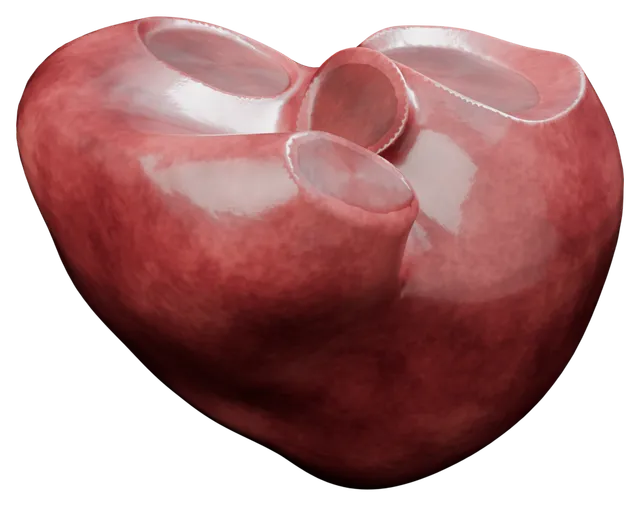
Drag to turn in 3D
One of the representative ventricular meshes (female_age_58_bmi_50), rendered from the published data, which are shared under CC0. Virtual Cohort of Four-Chamber Heart Meshes from Atrial Fibrillation Patient CT
2025
Four-chamber heart meshes built from end-diastolic CT of ten patients with atrial fibrillation, each in VTK and CARP formats. The post-processed versions add fibre directions in the atria and ventricles, fast endocardial conduction layers, Bachmann's bundle and an atrioventricular isolation surface.
Barrows RK, Augustin CM, Gsell MAF, Roney CH, Solís Lemus JA, et al. (2025) Rhythm control benefits left ventricular function compared with rate control in patients with atrial fibrillation: A computational study. Heart Rhythm O2 6(7): 1011-1019
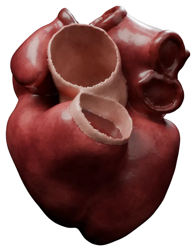
Drag to turn in 3D
Case 03 of the ten four-chamber meshes, rendered from the published data, which are shared under CC BY 4.0. Cardiac length-dependent activation driven by force-dependent thick-filament dynamics
2024
This study presents a model framework describing myocyte contraction dynamics that includes the effects of thick-filament regulation via the force-dependent crossbridge "off" state. The model builds upon the contraction model by Land et al. (2017) originating from this group.
Lewalle A, Milburn G, Campbell KS, Niederer SA. Cardiac length-dependent activation driven by force-dependent thick-filament dynamics. Biophysical Journal. 2024 Sep 17;123(18):2996-3009.
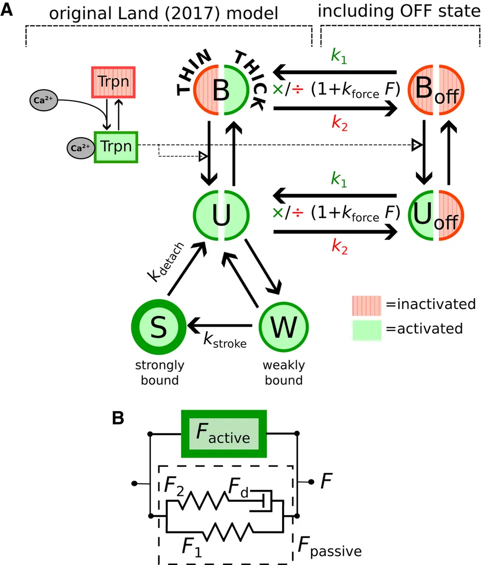
The kinetic scheme of the model, with the thick-filament OFF states B off and U off added to the states B, U, W and S of the Land model (A), and the active and passive forces of the sarcomere (B). Figure 1 of Lewalle et al. (2024), Biophysical Journal, under CC BY 4.0. Model Reproducibility Study on Left Atrial Fibres
2023
One hundred left atrial models built with CemrgApp by five operators from the contrast-enhanced MR angiograms and late gadolinium enhanced MRI of 50 patients: 60 for the variability between operators and 40 for the variability within one operator, with a file pairing the models compared. Each holds the labelled surface mesh, its fibres, and the local activation time and phase singularity maps of the pacing and atrial fibrillation simulations run on it in openCARP.
Solís-Lemus JA, Baptiste T, Barrows R, Sillett C, Gharaviri A, et al. (2023) Evaluation of an open-source pipeline to create patient-specific left atrial models: A reproducibility study. Comput Biol Med 162: 107009
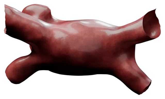
Drag to turn in 3D
Model M88 of the 100 left atrial models, rendered from the published data, which are shared under CC BY 4.0. Cell to Whole Organ Global Sensitivity Analysis on a Four-chamber Heart Electromechanics Model Using Gaussian Processes Emulators - Training Datasets
2023
This database contains all training datasets for the Gaussian processes emulators (GPEs) trained in the study entitled "Cell to Whole Organ Global Sensitivity Analysis on a Four-chamber Electromechanics Model Using Gaussian Processes Emulators", submitted to PLOS Computational Biology.
Strocchi M, Longobardi S, Augustin CM, Gsell MAF, Petras A, Rinaldi CA, et al. (2023) Cell to whole organ global sensitivity analysis on a four-chamber heart electromechanics model using Gaussian processes emulators. PLoS Comput Biol 19(6): e1011257. https://doi.org/10.1371/journal.pcbi.1011257
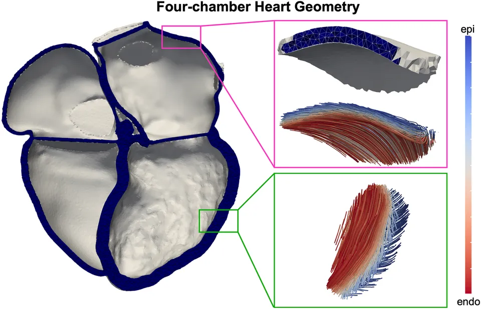
The four-chamber heart geometry, with fibre directions from endocardium to epicardium. Figure 1 of Strocchi et al. (2023), PLoS Computational Biology, under CC BY 4.0. Whole Torso Computational Models
2023
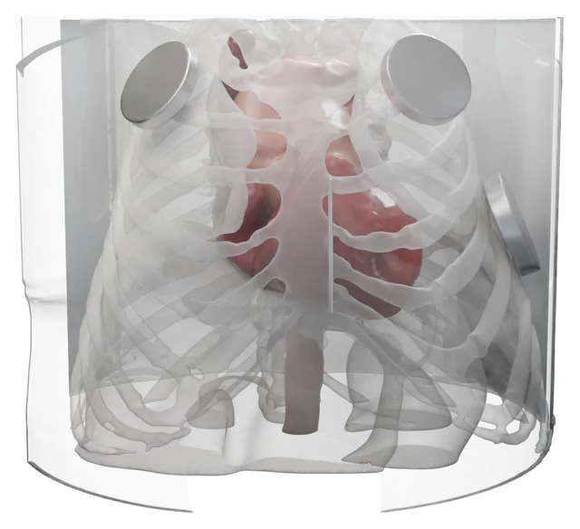
Whole torso computational models generated from high-resolution CT data.
Qian S, Monaci S, Mendonca-Costa C, Campos F, Gemmell P, Zaidi HA, Rajani R, Whitaker J, Rinaldi CA, Bishop MJ. Additional coils mitigate elevated defibrillation threshold in right-sided implantable cardioverter defibrillator generator placement: a simulation study. Europace. 2023 Jun;25(6):euad146.
Drag to turn in 3D
Torso 3 of the 5, with its organs, heart, great vessels and ICD leads and cans drawn inside the body, rendered from the published data, which are shared under CC BY 4.0. Constructing a Human Atrial Fibre Atlas
2021
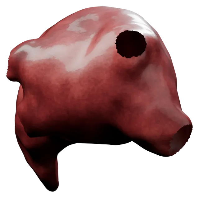
Endocardial and epicardial surfaces of the left and right atria from seven anatomies, each with its fibre field, and the atlas fibre field averaged across them, shown on the sixth anatomy. Every surface also carries universal atrial coordinates.
Roney, C.H., Bendikas, R., Pashakhanloo, F. et al. Constructing a Human Atrial Fibre Atlas. Ann Biomed Eng 49, 233–250 (2021). https://doi.org/10.1007/s10439-020-02525-w
Drag to turn in 3D
The epicardial surface of the left atrium that carries the atlas fibre field, rendered from the published data, which are shared under CC BY 4.0. Predicting Atrial Fibrillation Recurrence by Combining Population Data and Virtual Cohorts of Patient-Specific Left Atrial Models
2021
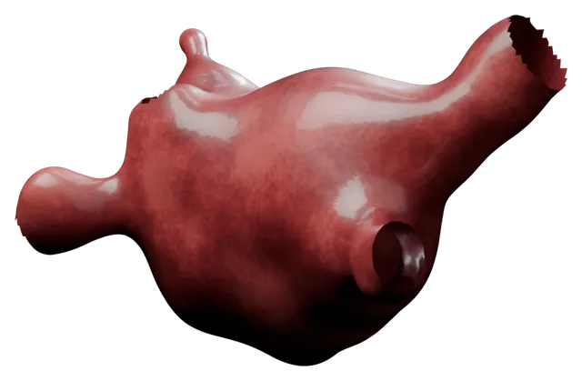
We include surface meshes in vtk format, consisting of the nodes, triangular elements, the atrial coordinate fields defined on the nodes, and the endocardial and epicardial fibre fields defined on the elements.
Roney, Caroline H., Iain Sim, Jin Yu, Marianne Beach, Arihant Mehta, Jose Alonso Solis-Lemus, Irum Kotadia et al. Predicting Atrial Fibrillation Recurrence by Combining Population Data and Virtual Cohorts of Patient-Specific Left Atrial Models. Circulation: Arrhythmia and Electrophysiology (2021): CIRCEP-121.
Drag to turn in 3D
One of the left atrial meshes (Mesh_83815278), rendered from the published data, which are shared under CC BY 4.0. Virtual cohort of 1000 synthetic heart meshes from adult human healthy population
2021
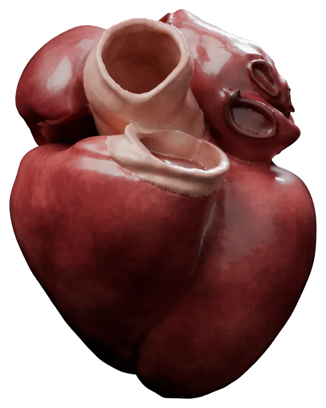
One thousand four-chamber heart models generated from a statistical shape model of the healthy CT cohort, for electromechanics simulations. The weights of the shape model are included.
Rodero C, Strocchi M, Marciniak M, Longobardi S, Whitaker J, O’Neill MD, Gillette K, Augustin C, Plank G, Vigmond EJ, Lamata P. Linking statistical shape models and simulated function in the healthy adult human heart. PLoS computational biology. 2021 Apr 15;17(4):e1008851.
Drag to turn in 3D
Synthetic heart 1 of the 1,000, rendered from the published data, which are shared under CC BY 4.0. Virtual cohort of adult healthy four-chamber heart meshes from CT images
2021
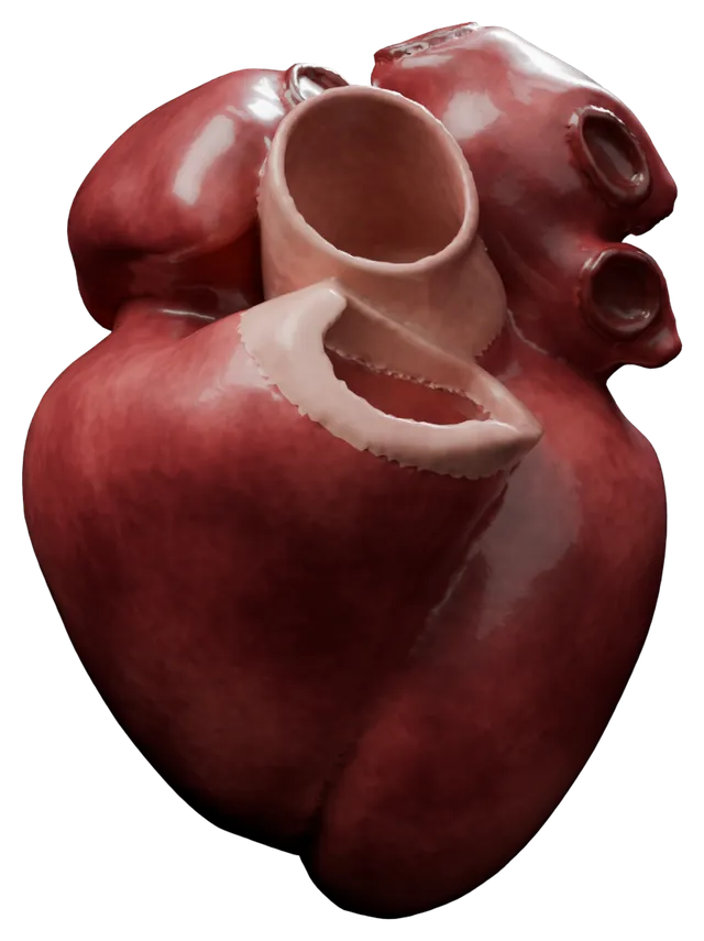
Twenty four-chamber heart models for electromechanics simulations, built from end-diastolic CT of patients who attended the emergency department with acute chest pain and in whom no cardiac condition was found at follow-up. The simulation outputs and the weights of the statistical shape model built from them are included.
Rodero C, Strocchi M, Marciniak M, Longobardi S, Whitaker J, O’Neill MD, Gillette K, Augustin C, Plank G, Vigmond EJ, Lamata P. Linking statistical shape models and simulated function in the healthy adult human heart. PLoS computational biology. 2021 Apr 15;17(4):e1008851.
Drag to turn in 3D
Case 18 of the 20 four-chamber meshes, rendered from the published data, which are shared under CC BY 4.0. Virtual cohort of extreme and average four-chamber heart meshes from statistical shape model
2021
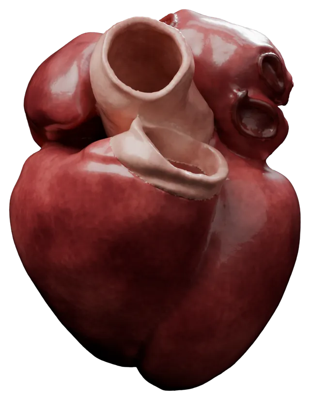
Thirty-nine four-chamber heart models from a statistical shape model of the healthy CT cohort: the average shape and shapes along its first nine modes, for electromechanics simulations. The simulation outputs and the weights of the shape model are included.
Rodero C, Strocchi M, Marciniak M, Longobardi S, Whitaker J, O’Neill MD, Gillette K, Augustin C, Plank G, Vigmond EJ, Lamata P. Linking statistical shape models and simulated function in the healthy adult human heart. PLoS computational biology. 2021 Apr 15;17(4):e1008851.
Drag to turn in 3D
The average shape of the statistical shape model, rendered from the published data, which are shared under CC BY 4.0. A publicly available virtual cohort of four-chamber heart meshes for cardiac electro-mechanics simulations
2020
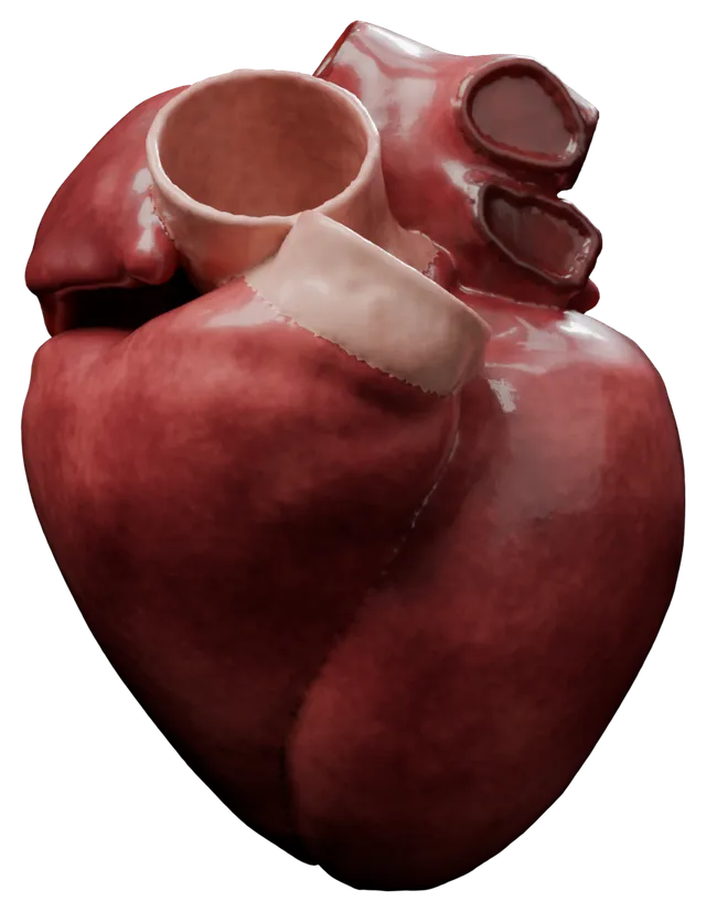
Twenty-four four-chamber heart models for electromechanics simulations, built from end-diastolic CT of heart failure patients recruited for an upgrade to cardiac resynchronisation therapy. Each mesh is also given at a higher resolution.
Strocchi M, Augustin CM, Gsell MAF, Karabelas E, Neic A, et al. (2020) A publicly available virtual cohort of four-chamber heart meshes for cardiac electro-mechanics simulations. PLOS ONE 15(6): e0235145
Drag to turn in 3D
Case 23 of the 24 four-chamber meshes, rendered from the published data, which are shared under CC BY 4.0. A Virtual Cohort of Twenty-four Left-ventricular Models of Ischemic Cardiomyopathy Patients
2020
Twenty-four left ventricular models of patients with ischaemic cardiomyopathy undergoing cardiac resynchronisation therapy, built from late gadolinium enhanced MRI, for electrophysiology simulations. Each includes the infarct scar core and border zone, rule-based fibres and universal ventricular coordinates.
Mendonca Costa, Caroline, Neic, Aurel Medical University of Graz, and Kerfoot, Eric King’s College London. A Virtual Cohort of Twenty-four Left-ventricular Models of Ischemic Cardiomyopathy Patients (2020)
Drag to turn in 3D
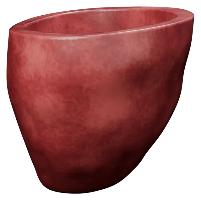
Case 4 of the 24 left ventricular models, rendered from the published data, which are shared under ODC-By. The Impact of Wall Thickness and Curvature on Wall Stress in Patient-Specific Electromechanical Models of the Left Atrium
2020
Finite element meshes of the left atrium from three patients, with rule-based myofibre directions. The third case is also given with Gaussian noise added to its geometry, and with a constant wall thickness of 0.5 mm.
Augustin CM, Fastl TE, Neic A, et al. The impact of wall thickness and curvature on wall stress in patient-specific electromechanical models of the left atrium. Biomech Model Mechanobiol. 2020;19(3):1015‐1034. doi:10.1007/s10237-019-01268-5
Drag to turn in 3D
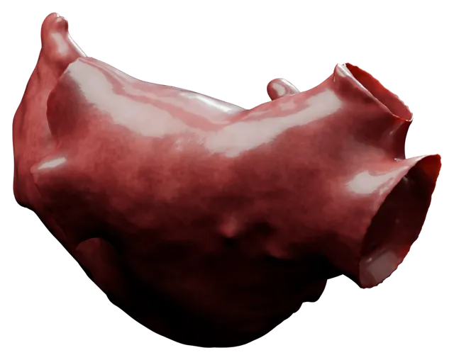
The left atrial mesh of patient 3, rendered from the published data, which are shared under CC BY 4.0. Personalized computational finite element models for left atrial electromechanics
2018
Finite element models of the left atrium from coronary CT angiography of three patients at Guy's and St Thomas' NHS Foundation Trust, for electromechanics simulations. Each has a high-resolution tetrahedral mesh and a myofibre architecture estimated from anatomical data.
Fastl, Thomas E, Tobon-Gomez, Catalina King's College London, Crozier, Andrew Medical University of Graz, and McCarthy, Karen P Royal Brompton Hospital. Personalized Computational Finite Element Models for Left Atrial Electromechanics (2018).
Drag to turn in 3D
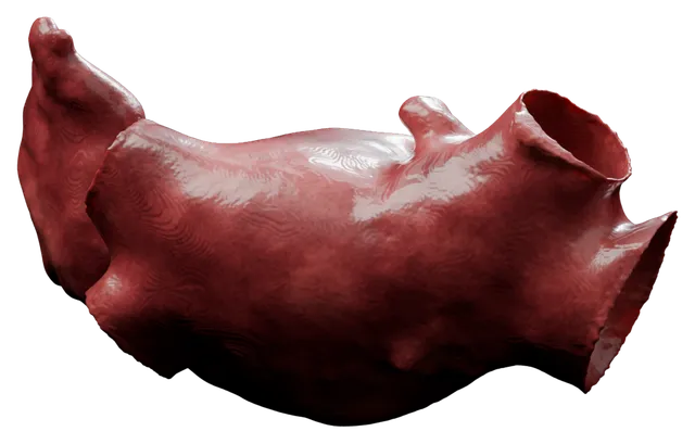
The left atrial model of patient 3, rendered from the published data, which are shared under CC BY-NC-SA 4.0, as this image is. A model of cardiac contraction based on novel measurements of tension development in human cardiomyocytes.
2017
A model of human cardiac contraction intended for multi-scale modelling applications. Download includes relevant experimental data, and C++ code is available on request.
S Land, SJ Park-Holohan, NP Smith, CG Dos Remedios, JC Kentish, SA Niederer. JMCC, 2017
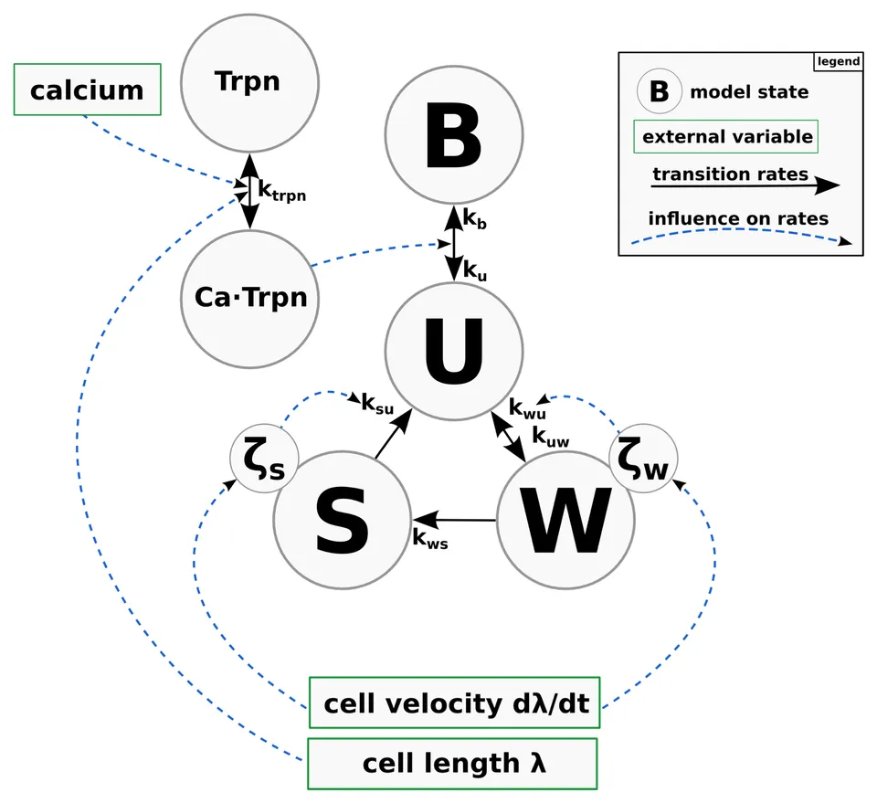
The active tension model: the blocked state B, the unbound crossbridge state U, the pre-powerstroke state W and the force-generating state S, with ζw and ζs tracking the mean distortion of the crossbridges in W and S. Calcium binding to troponin C (Trpn) drives the transition between B and U. Figure 3 of Land et al. (2017), Journal of Molecular and Cellular Cardiology, from the accepted manuscript, under CC BY-NC-ND 4.0. A Biophysical Systems Approach to Identifying the Pathways of Acute and Chronic Doxorubicin Mitochondrial Cardiotoxicity
2016
The clinical use of the anthracycline doxorubicin is limited by its cardiotoxicity which is associated with mitochondrial dysfunction. Redox cycling, mitochondrial DNA damage and electron transport chain inhibition have been identified as potential mechanisms of toxicity. However, the relative roles of each of these proposed mechanisms are still not fully understood. The purpose of this study is to identify which of these pathways independently or in combination are responsible for doxorubicin toxicity. A state of the art mathematical model of the mitochondria including the citric acid cycle, electron transport chain and ROS production and scavenging systems was extended by incorporating a novel representation for mitochondrial DNA damage and repair. In silico experiments were performed to quantify the contributions of each of the toxicity mechanisms to mitochondrial dysfunction during the acute and chronic stages of toxicity. Simulations predict that redox cycling has a minor role in doxorubicin cardiotoxicity. Electron transport chain inhibition is the main pathway for acute toxicity for supratherapeutic doses, being lethal at mitochondrial concentrations higher than 200μM. Direct mitochondrial DNA damage is the principal pathway of chronic cardiotoxicity for therapeutic doses, leading to a progressive and irreversible long term mitochondrial dysfunction.
de Oliveira BL, Niederer S. A biophysical systems approach to identifying the pathways of acute and chronic doxorubicin mitochondrial cardiotoxicity. PLoS Computational Biology. 2016 Nov 21;12(11):e1005214.
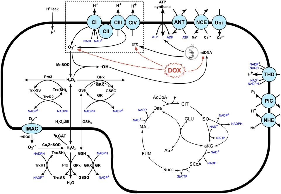
The mitochondrial model, with the sites where doxorubicin (DOX) acts. Figure 7 of de Oliveira and Niederer (2016), PLoS Computational Biology, under CC BY 4.0. A Spatially Detailed Model of Isometric Contraction Based on Competitive Binding of Troponin I Explains Cooperative Interactions between Tropomyosin and Crossbridges
2015
A spatially detailed ODE model of contraction based on continuous flexible chain model of the thin filament. Includes explicit TnI kinetics and a biophysically detailed representation of RU-RU, RU-XB, and XB-XB cooperativity.
Land S, Niederer SA. PLoS Computational Biology, 2015
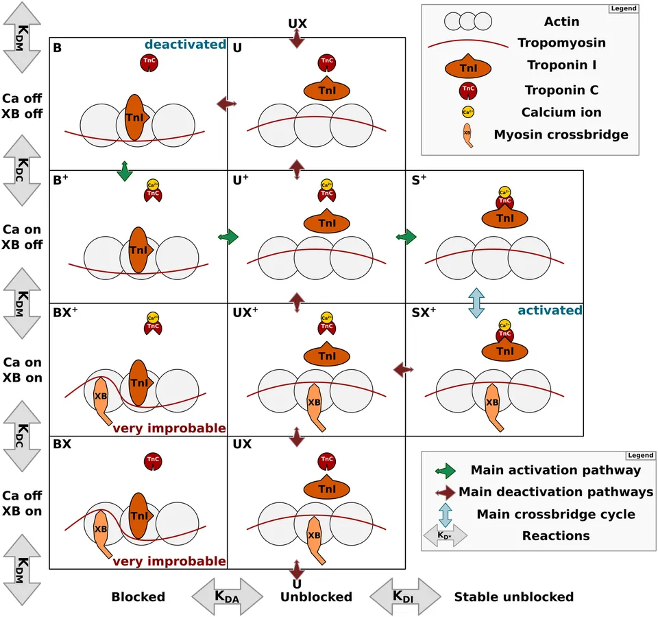
The states of a regulatory unit, from blocked to stable unblocked, with troponin I, troponin C, tropomyosin and the myosin crossbridge. Figure 1 of Land and Niederer (2015), PLoS Computational Biology, under CC BY 4.0. Verification of cardiac mechanics software: benchmark problems and solutions for testing active and passive material behaviour
2015
To aid in the verification of current and future cardiac mechanics solvers, this study provides three benchmark problems for cardiac mechanics. These benchmark problems test the ability to accurately simulate pressure-type forces that depend on the deformed objects geometry, anisotropic and spatially varying material properties similar to those seen in the left ventricle and active contractile forces.
Land S, Gurev V, Arens S, Augustin CM, Baron L, Blake R, Bradley C, Castro S, Crozier A, Favino M, Fastl TE. Verification of cardiac mechanics software: benchmark problems and solutions for testing active and passive material behaviour. Proceedings of the Royal Society A: Mathematical, Physical and Engineering Sciences. 2015 Dec 8;471(2184):20150641.
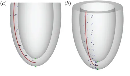
The reference geometry of the ventricle in benchmark problems 2 and 3. Panels a and b of Figure 2 of Land et al. (2015), Proceedings of the Royal Society A, under CC BY 4.0. An analysis of deformation-dependent electromechanical coupling in the mouse heart
2012
A model of contraction fitted to mouse data at physiological temperature and frequency, including length and velocity dependent tension generation.
Land S, Niederer SA, Aronsen JM, Espe EK, Zhang L, Louch WE, Sjaastad I, Sejersted OM, Smith NP. J Physiol. 2012 15;590(Pt 18):4553-69. Epub 2012 May 21. PubMed ID: 22615436
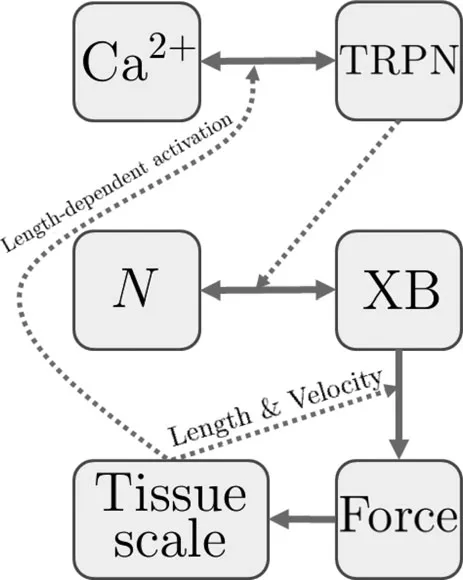
The cell model: calcium binds cooperatively to troponin C (TRPN), which increases the fraction of actively cycling crossbridges (XB), and length and velocity dependencies give the force used at the tissue scale. Diagram from the model's entry in the Physiome Model Repository, under CC BY 3.0. A mathematical model of the murine ventricular myocyte: a data-driven biophysically based approach applied to mice overexpressing the canine NCX isoform
2010
A mouse electrophysiology model fitted to mouse data at physiological temperatures and capable of pacing to physiological pacing rates.
L. Li , S. A. Niederer , W. Idigo , Y. H. Zhang , P. Swietach , B. Casadei , N. P. Smith, 2010 American Journal of Physiology, Volume 299 no. H1045-H1063
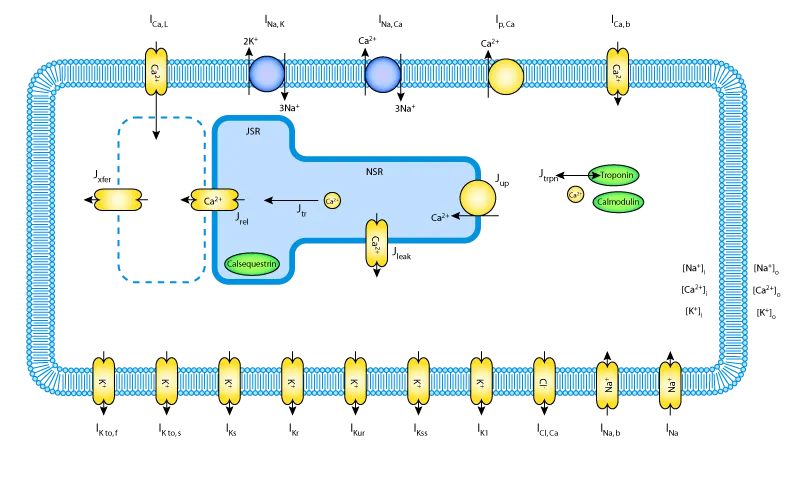
The mouse ventricular myocyte model of Bondarenko et al. (2004), into which this model's calcium handling components were incorporated. Diagram from the model's entry in the Physiome Model Repository, under CC BY 3.0. A Quantitative Analysis of Cardiac Myocyte Relaxation: A Simulation Study
2006
A biophysical data driven model of length, velocity and calcium dependent tension development in rat cardiac muscle at room temperature.
Steven Niederer, Peter Hunter, Nicholas Smith, 2006 Biophysical Journal, 90 1697-1722 PubMed ID: 16339881
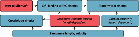
The active contraction framework proposed by Hunter et al., on which the model builds. Diagram from the model's entry in the Physiome Model Repository, under CC BY 3.0.