Imaging, anatomy and shape
Turning clinical images into anatomical models: segmentation, meshing, fibre architecture and statistical shape analysis, at the scale of whole cohorts rather than single cases.
Advancing the understanding, diagnosis and treatment of heart disease through physiology-based simulation and machine learning.
Its 25 published models and 6 software tools are open for other groups to use.
Join us
Figures from the group's work, from a single cell to a cohort of patients. Each one opens the research theme it belongs to.
Cell
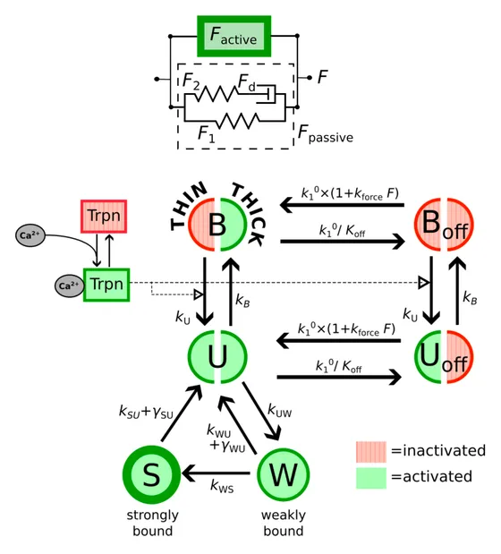
A myocyte-level contraction model featuring the myosin cross-bridge “off” state, proposed as an underlying mechanism that regulates the dependence of generated force on the sarcomere length in cardiac muscle.
Adapted from Figure 1 of Lewalle et al. (2024), Biophysical Journal, CC BY 4.0
Cells, drugs and safety
Body
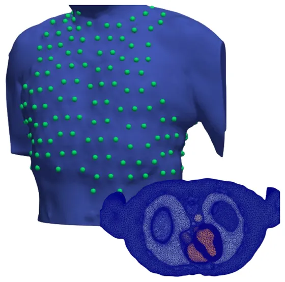
A torso mesh built from CT, with the electrode positions of a 252-lead vest, used to simulate body surface potentials.
Figure 1 of Malik et al. (2026), PLOS Computational Biology, CC BY 4.0
Ventricular arrhythmia and risk
Cohort
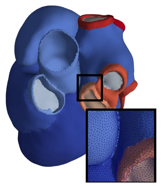
Four-chamber heart models of 25 men and 25 women, with heart failure and without, released as a public virtual cohort.
Figure 3 of Solís-Lemus, Barrows et al. (2026), PLOS Computational Biology, CC0
Digital twins and virtual cohorts
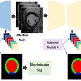
Turning clinical images into anatomical models: segmentation, meshing, fibre architecture and statistical shape analysis, at the scale of whole cohorts rather than single cases.
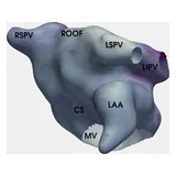
Mechanisms that start and sustain atrial fibrillation, which patients an ablation will help, and how it can be targeted for a particular atrium rather than a generic one.
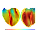
Why dangerous ventricular rhythms arise in scarred and remodelled hearts, which regions of a particular heart can sustain them, and where an ablation should be aimed.
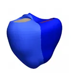
Models tied to one person's measurements, built for many people at once, so that questions can be asked of a population of hearts rather than a single example.
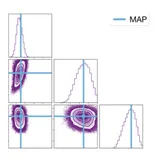
How far a model's predictions can be trusted: emulators, sensitivity analysis and calibration that state what the data can identify, and verification of the simulators themselves.
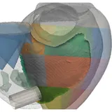
Electromechanical models of the failing and hypertrophied heart, and how pacing, resynchronisation and other treatments can be planned for a particular patient.
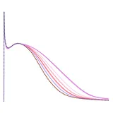
Biophysical models of the myocyte, from ion channels to force generation, applied to drug action and safety, to sex hormones, and to carrying results from the laboratory to patients.
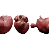
The group's 16 public collections of cardiac and torso meshes, shared on Zenodo with its collaborators, now hold more than 2,700 models and have been downloaded over 88,000 times.

Steven Niederer is on the organising committee of the 14th conference on Functional Imaging and Modelling of the Heart, FIMH 2027, to be held from 8 to 11 June 2027 at the London Institute for Healthcare Engineering.
A second week of teaching on cardiac digital twins, from 5 to 9 October 2026 at the Alan Turing Institute, free to attend and run through CVD-Net. Applications have closed.
The group's work is supported by the following funders, listed in order of how often they are named on its recent papers.

This project has received funding from the European Research Council (ERC) under the European Union's Horizon 2020 research and innovation programme (Grant agreement No. 864055).
This research was supported by the NIHR Imperial Biomedical Research Centre. The views expressed are those of the author(s) and not necessarily those of the NIHR or the Department of Health and Social Care.
Nothing is open at the moment. Positions are advertised on the Join us page as soon as they open. For anything else, see Contact.